
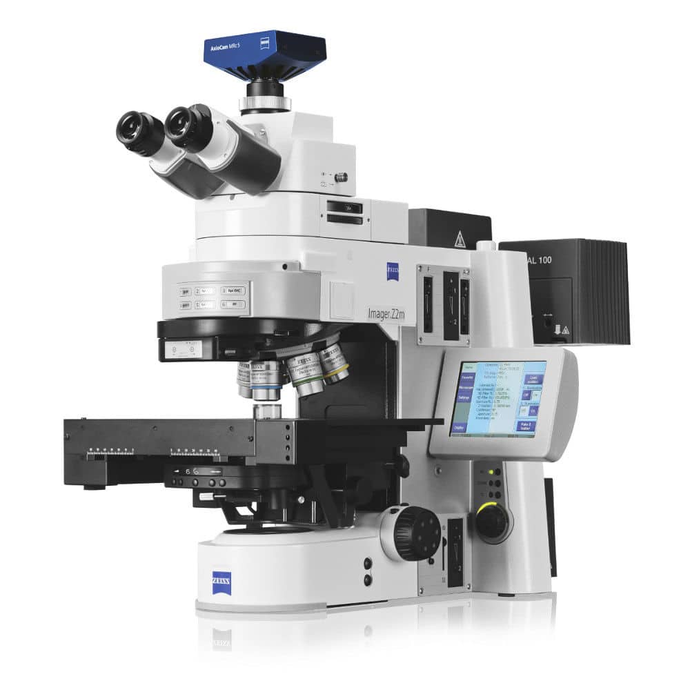
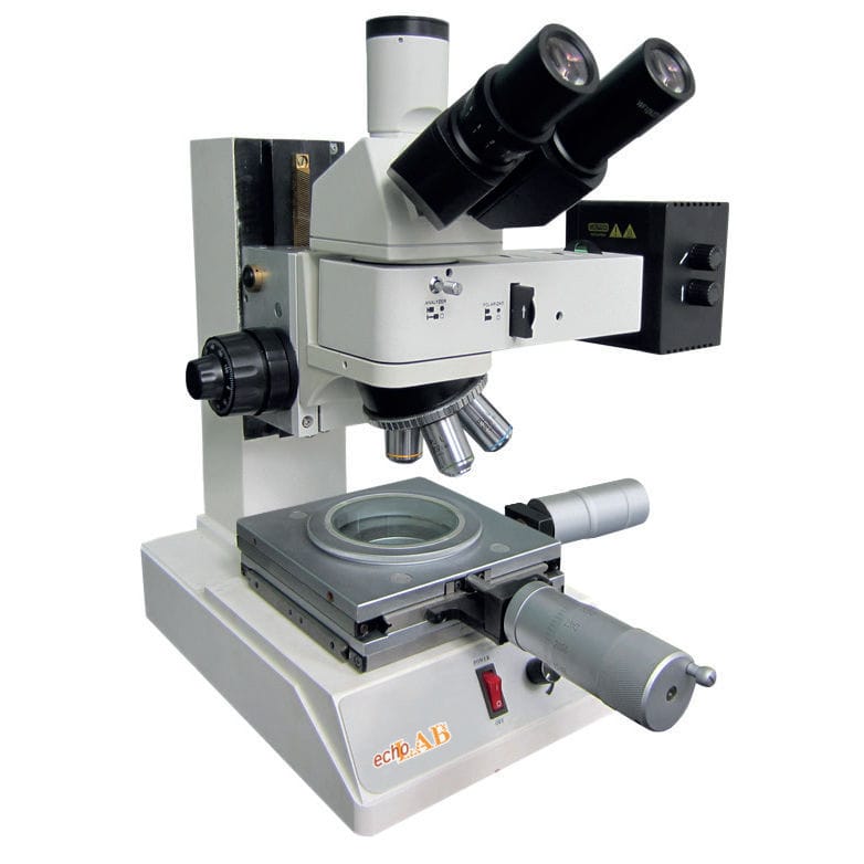
Carl Zeiss Microscopy optical microscope
There are two main types of microscopes: optical microscopes and electron microscopes.
The main difference between these two types of microscopes lies in the way the sample to be observed is prepared and passed through. This is what determines the quality of the image (magnification, color, black and white).
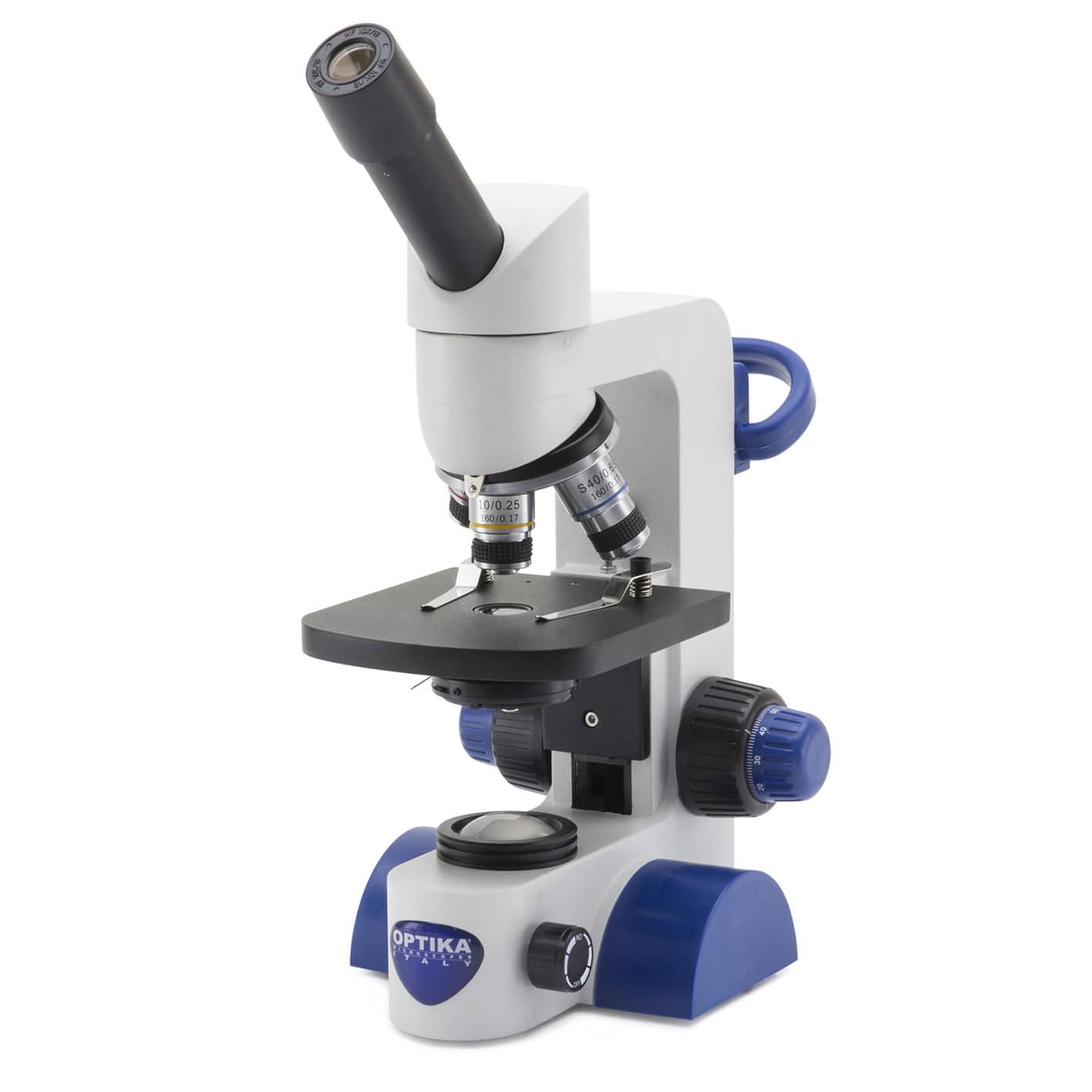
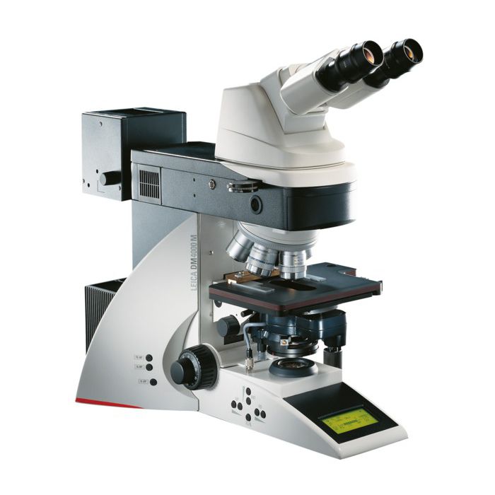
With an optical microscope, the preparation is placed on a glass slide and irradiated with light rays:
- The resolution is in the range of 200 nanometers.
- For example, it is possible to observe an entire cell.
- However, the magnification is rather limited, meaning that details smaller than 200 nanometers cannot be observed.
With an electron microscope, an electron beam is passed through the prepared sample:
- The magnification is higher.
- However, the image is in black and white.
- Colors can be added digitally on the computer afterwards.

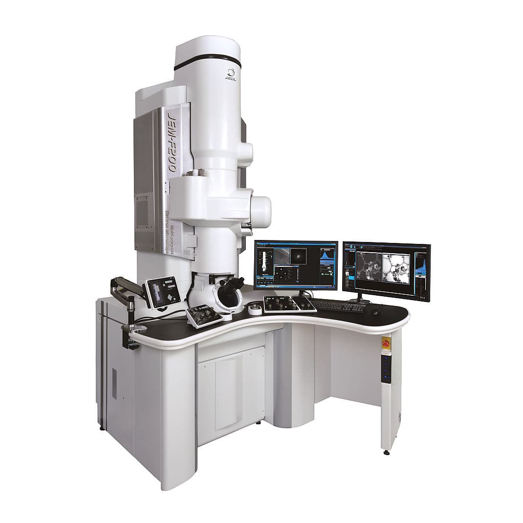
Jonel electron microscope
There are two types of electron microscopes: scanning and transmission.
- Scanning Electron Microscope (SEM):
- This type of microscope emits electrons that sweep the surface of the prepared sample.
- The resolution is very high, in the range of 0.4 to 20 nanometers, which makes it possible to differentiate between two points less than a nanometer apart.
- The relief image enables the structure and the form of the sample to be studied.
- This type of microscope is mainly used by biological research institutes to obtain the shape of cells or organs.
- They can cost between 150,000 and a million euros.
- Transmission Electron Microscope (TEM):
- This type of microscope emits electrons that go through the prepared sample.
- This means the resulting image provides even the most minute details of the sample.
- This type of microscope is used in cell biology because it is the only way to obtain accurate images of the inside of a cell.


Thank you for your sharing.